APPLICATIONS BY SPECIALTY
PreXion’s Versatility of Applications
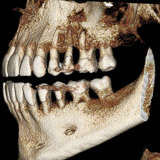
Among the many benefits of PreXion CBCT is the versatility of models’ applications. As part of our mission to Make IT Visible, we take great pride in producing technology that elevates dentistry developing superior image quality to serve many specialties. Below are just a few of the fields currently being empowered by PreXion 3D imaging.
Endodontics
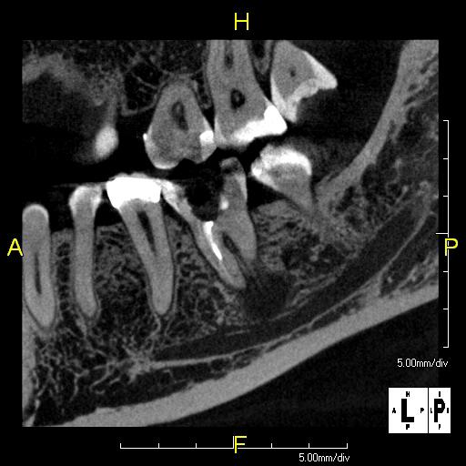
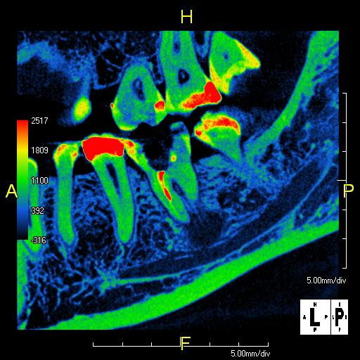
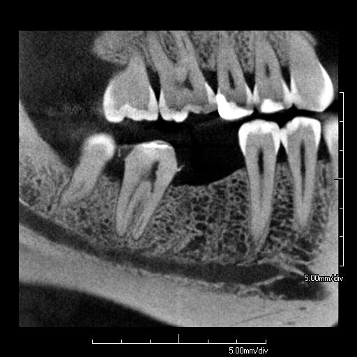
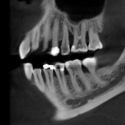
One of the greatest benefits of PreXion technology is the abundance of information provided by our scans. The cone beam captures a substantial number of sliced images, compiling them into more thorough 3D images. These provide a much clearer representation of the patient’s anatomy compared to conventional 2D X-ray methods. This can help reduce or eliminate endodontic retreatment rates by having more complete information. This more thorough picture can also assist in determining the exact locations of potential second mesial-buccal canals prior to surgeries, allowing at least a moderate simplification of a complex process.
Our powerful imaging technology also helps simplify clinical diagnoses across the board, including:
- Vertical root fracture
- Retained root tips
- Apical radiolucency
- Periapical pathology
- Dentoalveolar trauma
- Resorption (internal & external)


Implant Dentistry
The 3D imaging capabilities have many benefits, including simplified reorientation. Due to the isotropic nature of the volumetric data set, the accompanying software allows users to reorient in all three reference planes, meaning that the implant can be properly aligned into the patient’s bone using the optimal size for integration. CBCT imaging and software also improve reformatting and display ability. According to a clinical article published by Implant Dentistry U.S., “A CBCT image can be reformatted to panoramic, cephalometric, or bilateral multiplanar projections of the temporomandibular joint. These images, in turn, can be annotated, assessed, and measured for diagnostic and treatment planning purposes.”
Below is a list of additional benefits and applications, allowing clinicians to:
- Make measurements with an exact 1:1 ratio absent of superimposed structures or magnification
- Assess detailed bone quality and quantity and quickly determine if grafting is required
- Eliminate contingency treatment plans and surprises during implant surgery due to the ability to view hard and soft tissue structures, including the inferior alveolar nerve
- Lessen surgical trauma or exploratory surgery for patients as a result of more accurate pre-surgical measurements
- Create an optimal surgical guide fit
- Improve implant site selection and more easily predict prosthetic outcomes
- Increase production through improved case acceptance to quickly recoup costs and realize profits faster


Prosthodontics
PreXion CBCT scanners greatly improve treatment planning, allowing the specialist to visualize next steps in fully realized detail. According to a study published in the International Journal of Contemporary Medical Research (IJCMR), “CBCT provides a unique imaging option for various treatment needs of a prosthodontist. It can prove to be beneficial in various aspects of prosthodontic practice [e.g.,] from imaging of the temporomandibular joint for accurate movement simulation, to denture therapy. CBCT could play an important role in reduction of hectic routine for the clinician.”
Below are a few of the more granular applications of our technology in prosthodontics, which allow clinicians to:
- Easily export DICOM scans to third-party CAD/CAM software for complete treatment planning
- Integrate with all DICOM compliant third-party implant surgical planning software based on preference
- Plan complex implant cases (i.e. all on “4”) more precisely with superior image clarity and resolution
- Streamline implant case workflow in your practice
Periodontics
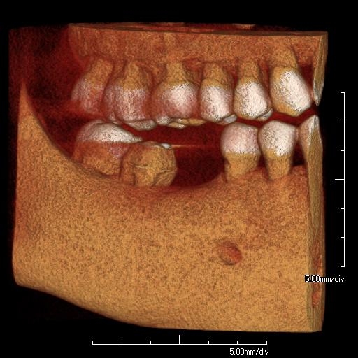
PreXion technology provides unmatched imaging clarity due to its departure from traditional imaging methodology. Unlike conventional CT scans, CBCT creates a fully 3-dimensional image by capturing hundreds of image “slices” of the patient and reconstructing them into a fully movable 3D rendering. Not only does it provide a groundbreaking level of clarity, but it also requires shorter scan times with less radiation exposure and yields an image that functions much more as a patient education tool than does a traditional static, 2D image. In addition, the contrast between bone and soft tissue optimizes diagnostic capability and periodontal treatment planning.
Our technology has extensive applications in periodontology, such as:
- Furcation involvement
- Soft tissue assessment
- Periodontal ligament space
- Alveolar bone defects
- Regenerative periodontal therapy
- Bone grafts


What is the Industry Saying?
What is the Industry Saying?
“WIth PreXion’s image quality, software capability, and customer support I can’t imagine anyone not using the PREXION CBCT who has an implant practice. The peace of mind and confidence this brings to every care - from the most simple to the most complex - is priceless.”